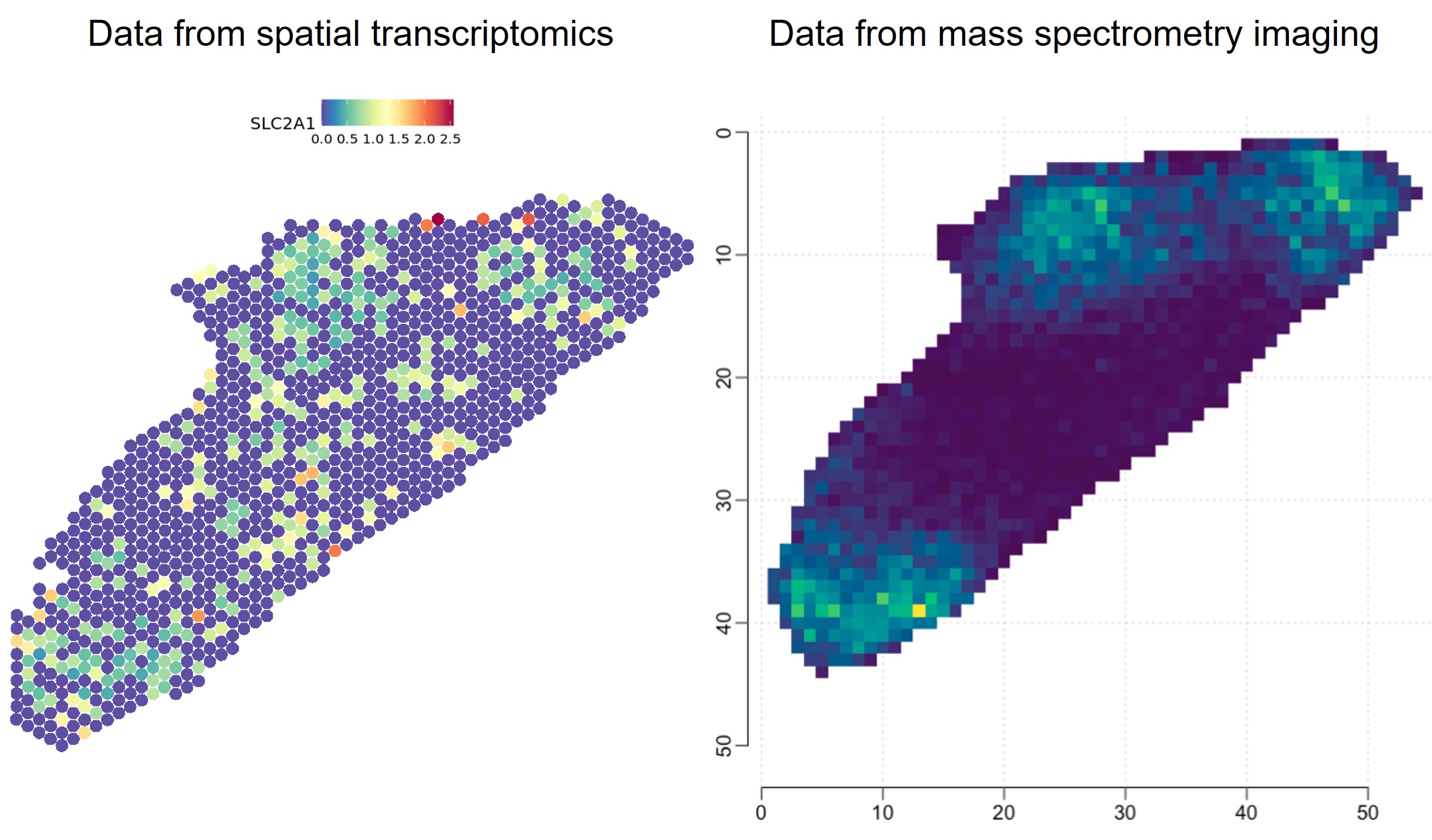
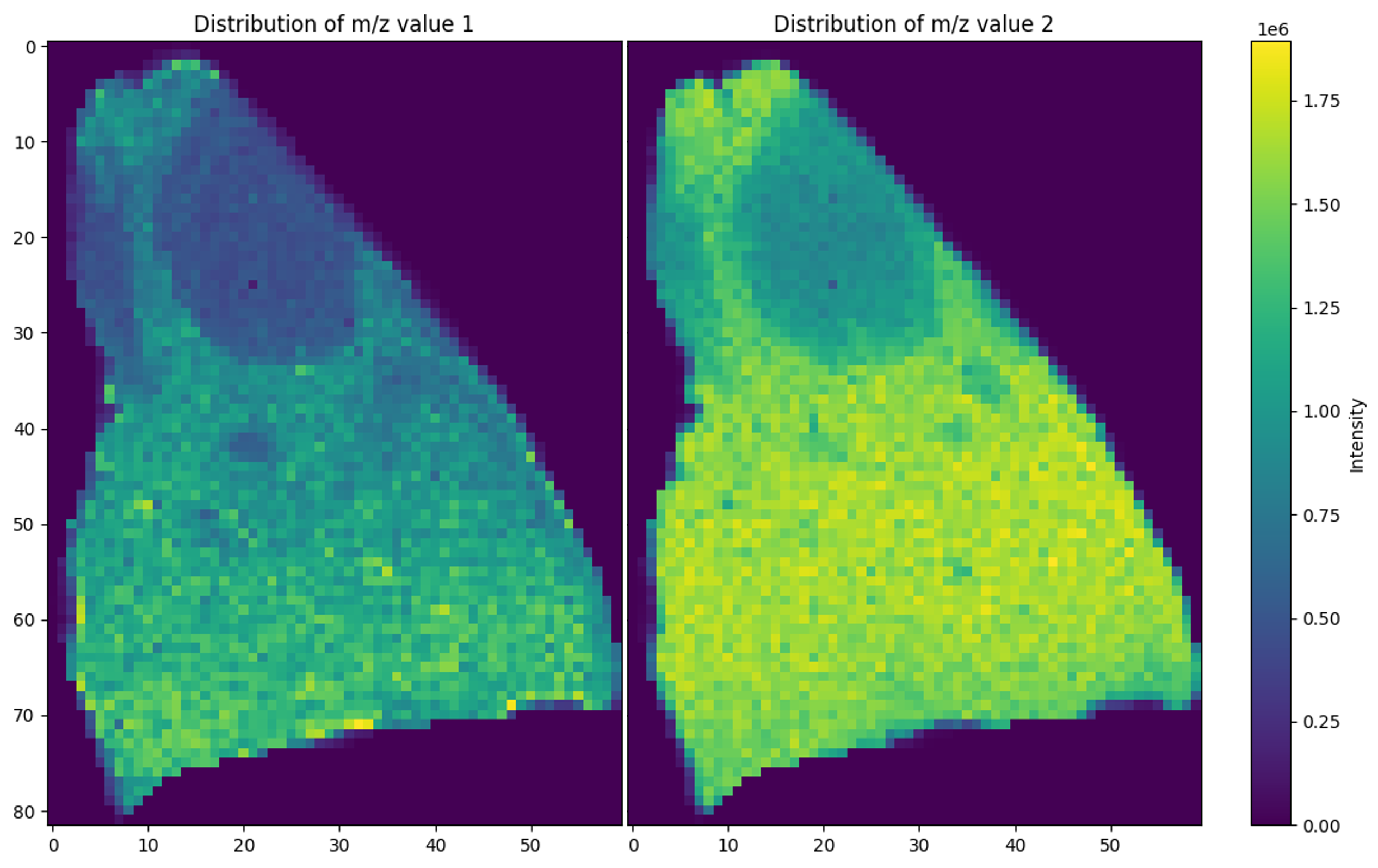
Combining data from mass spectrometry imaging and spatially resolved transcriptomics modalities
The project aims to integrate data from mass spectrometry imaging (MALDI MSI), spatial transcriptomics (Visium) and spatial VDJ and visualize them in TissUUmaps
Collaboration with Advanced Light Microscopy Facility
Collaboration between BIIF and ALM to produce tools for MINFLUX software
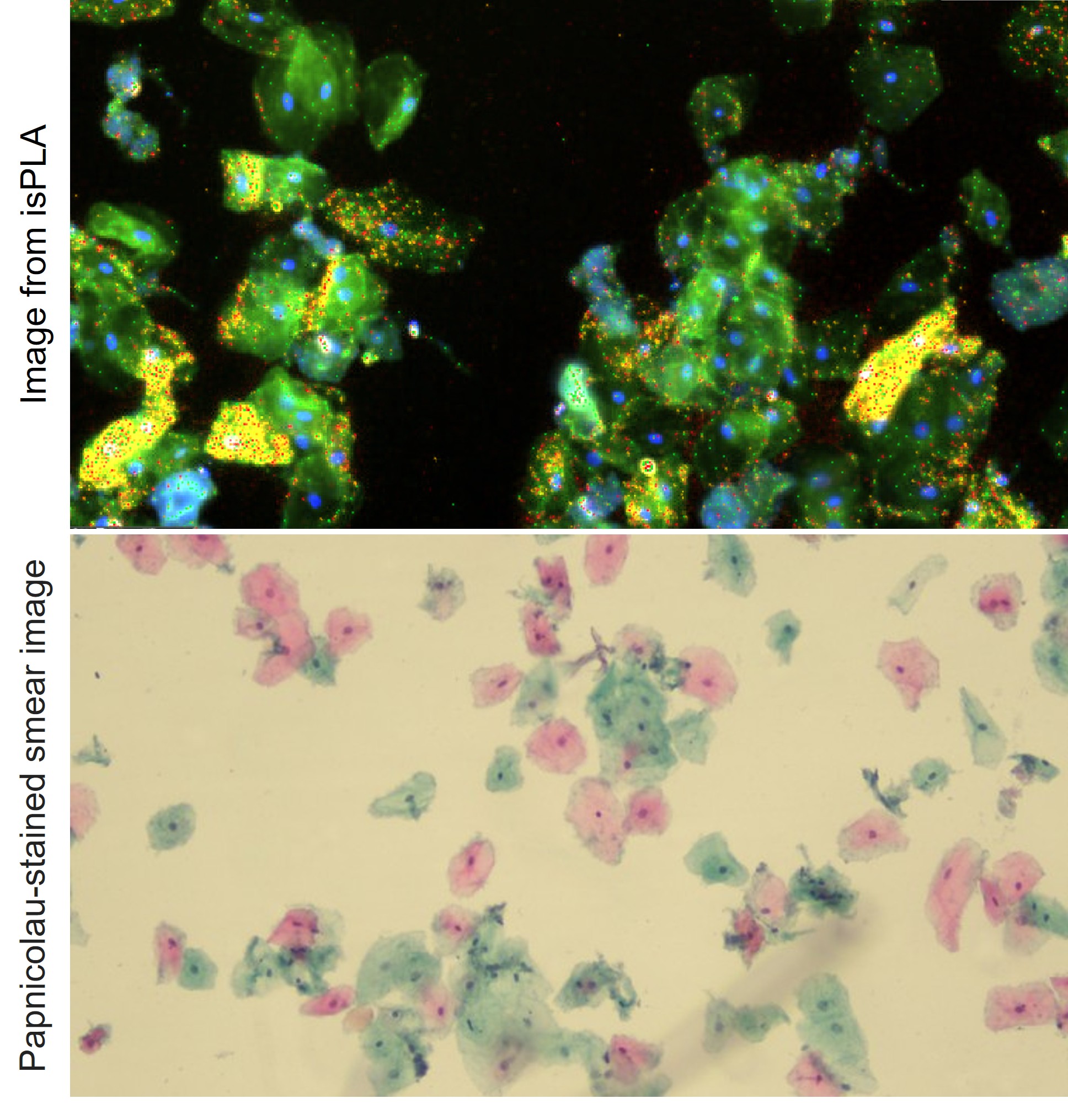
Early diagnostic of dysplastic cells from oral cavity
The incidence of oral cancer is increasing in the world and using a cytobrush to obtain a couple of thousand cells extracted from the abnormal area, typically located in the mucosa or gingiva,...
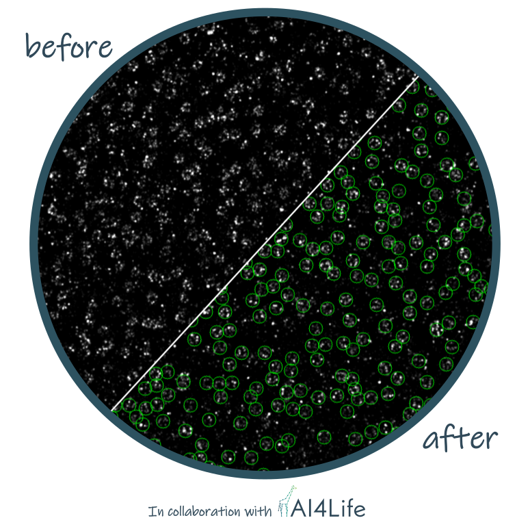
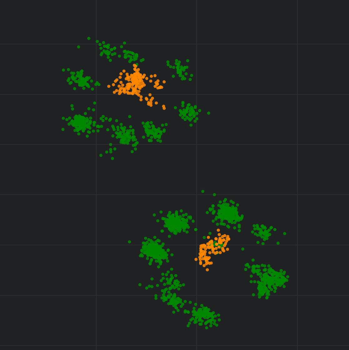
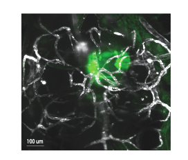
Detection of Nuclear Pore Complexes by DNA PAINT
The project’s aim was to identify nuclear pore complexes in DNA-PAINT images, including those that deviate from the canonical structure in terms of diameter, stoichiometry, and circularity.
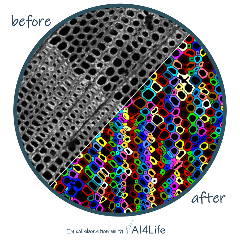
Imprints of Wind Disturbances on Wood Anatomy
This project investigates differences in wood quality among trees from the Boubín Forest nature reserve, the largest indigenous forest in Central Europe.
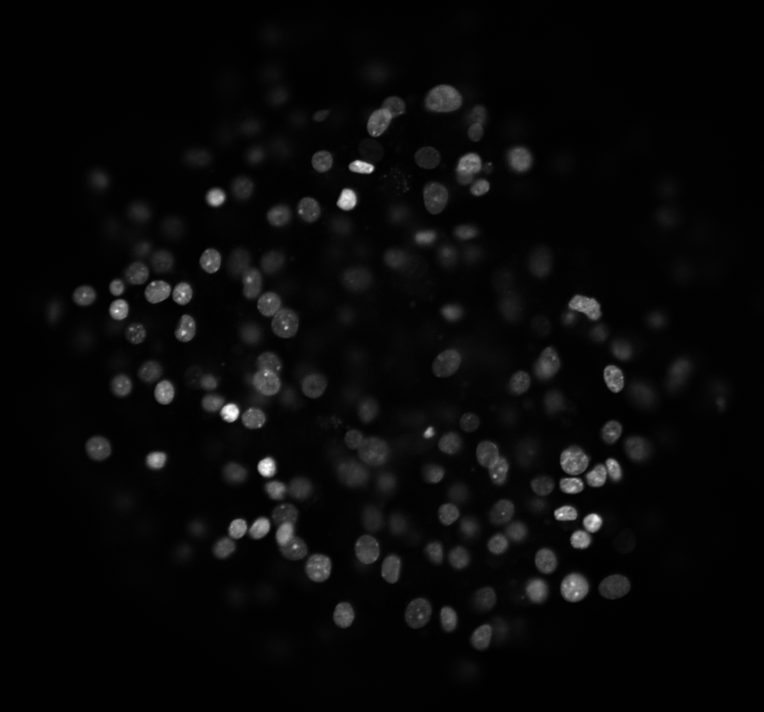
Amyloid Formation Under Stress Conditions in Pancreatic Islets
Pancreatic islet beta-cells are known to produce amyloid aggregates in response to various stress factors, including prolonged exposure to high glucose levels. These amyloid deposits are implicated in beta-cell dysfunction and the progression...
TDP - Spatial Biology
Integration of MSI and spatial proteomics data
Spatial Profiling of the renal host-pathogen interplay in response to Candida albicans infection...
We have used intra vital 2-photon microscopy combined with a customized spatial transcriptomics approach to study the virulence properties of the human fungal pathogen Candida albicans. Using mice as models, we have imaged...
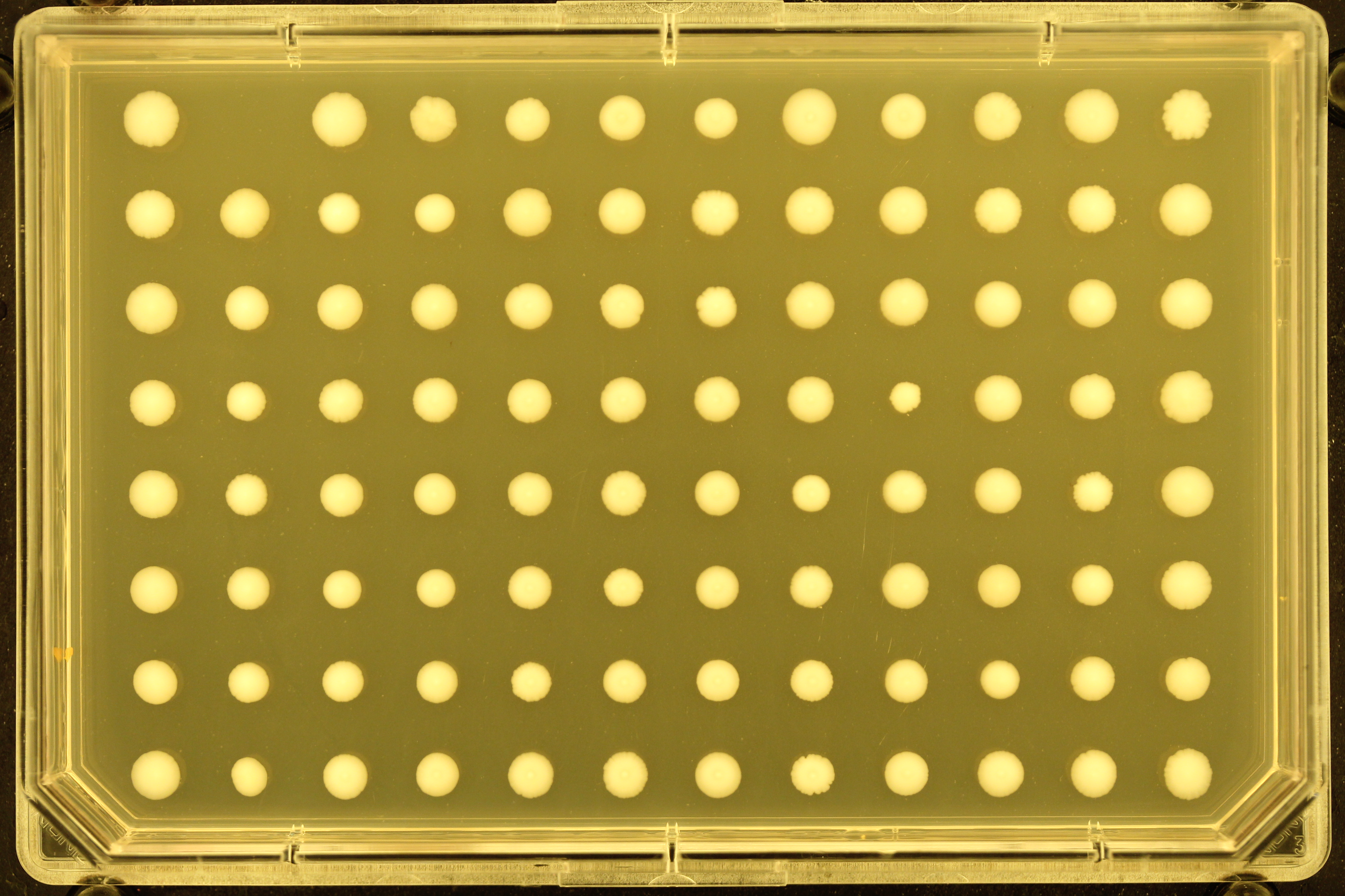
Automated Yeast Colony Quantification for Fatty Acid Sensitivity Screening
This project aims to develop an automated image analysis pipeline for a genome-wide yeast screen to identify mutants sensitive to fatty acids. The screen involves spotting yeast mutant arrays on media containing different types of...
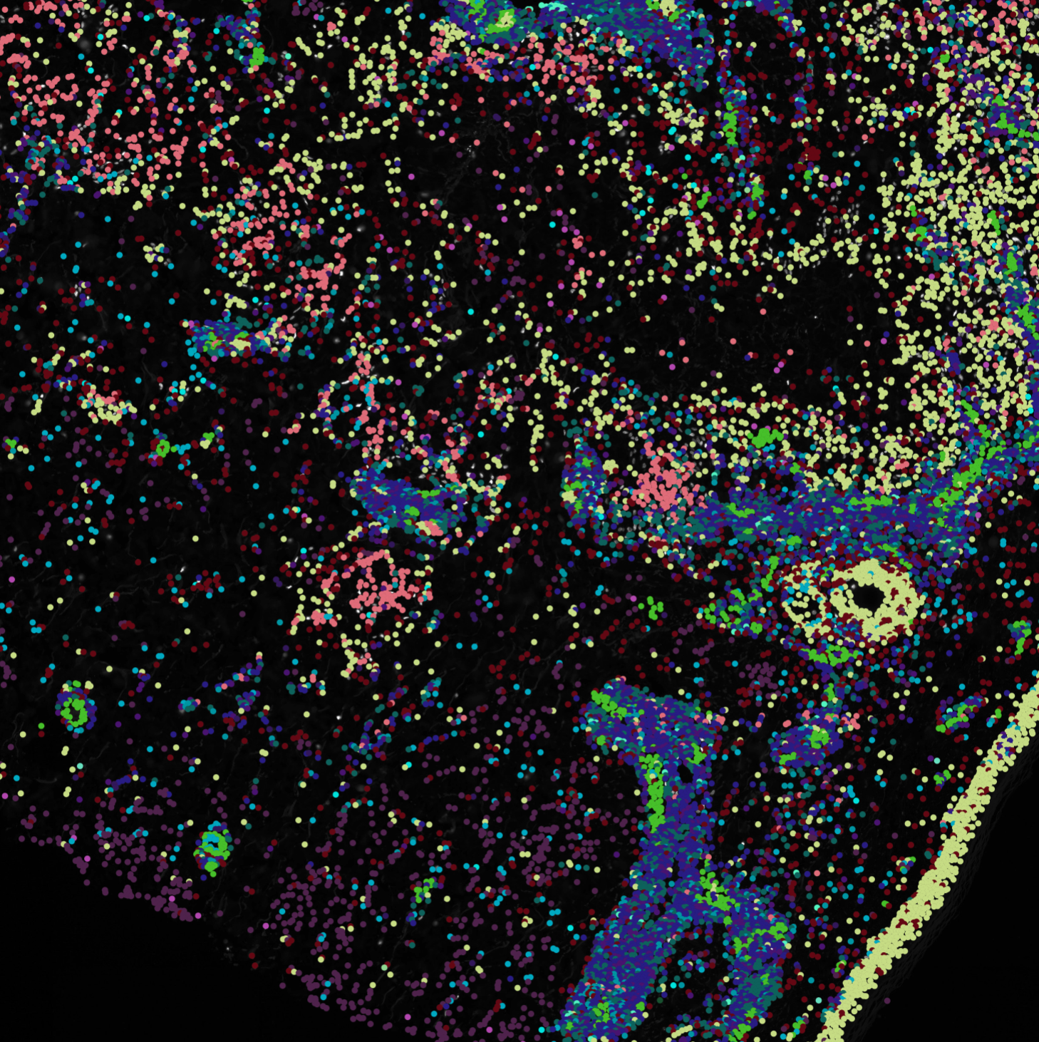
Phenocycler analysis of immune profile differences in granulomatous inflammation
Exploring whether the immune profile differ between Non-tuberculous mycobacteria and other granulomatous inflammations such as Granuloma Annulare and Sarcoidosis.
Cell dynamics of different glioblastoma invasion patterns in zebrafish xenografts
In this project, we are creating zebrafish xenografts by transplanting glioblastoma patient-derived cell lines tagged with GFP into Tg(kdrl:mcherry) zebrafish embryos.
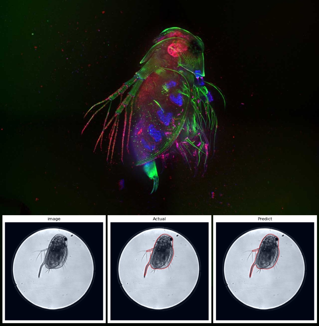
High-Throughput Analysis for Toxicological Assessment in Daphnia magna
This project aims to develop an image-based analysis method for toxicological effects in Daphnia magna, an ecotoxicological standard test species, after exposure to industrial chemicals or environmental samples. Molecular staining combined with fluorescence microscopy measures...
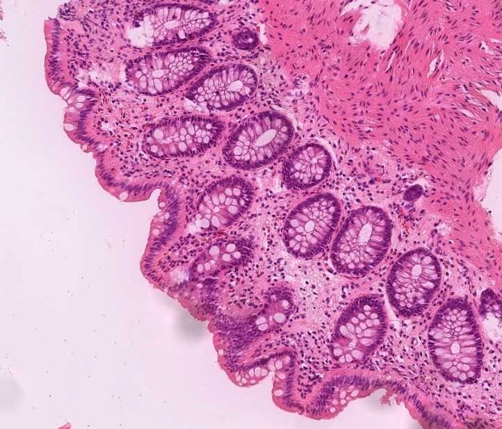
Gastrointestinal tissue-samples from population-based endoscopy studies
The project consists of gastrointestinal tissue-samples from 3 population-based endoscopy studies. In all three studies a random selection from the general population was included. In the Kalixanda upper endoscopy study includes biopsies from 1000 individuals,...
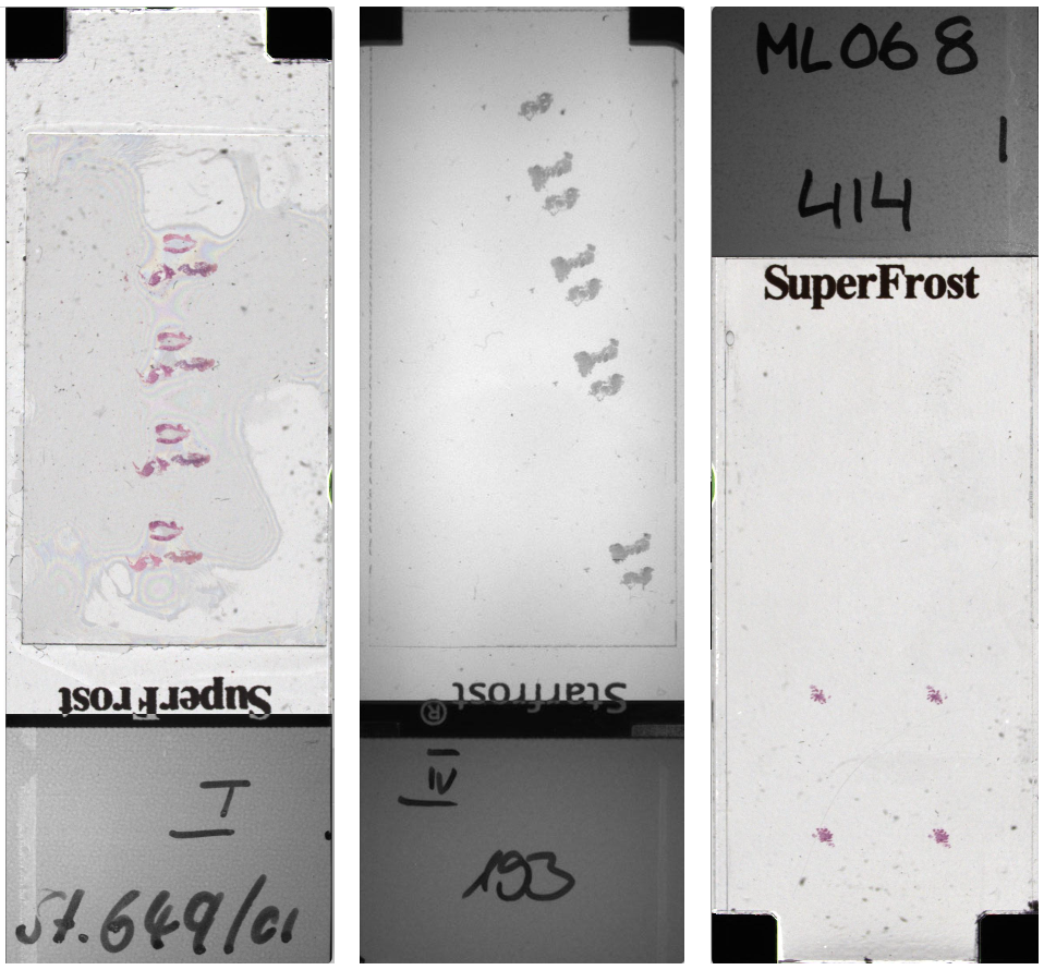
LLM-Accelerated Automated Handwritten Text Extraction from Microscopy Slides
The aim of this project is to structure the digitalized samples and to automatically extract the label id, year, marker information and slide number of the microscopy slides. We have used Qwen LLM...
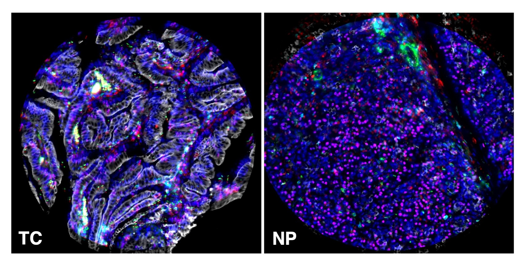
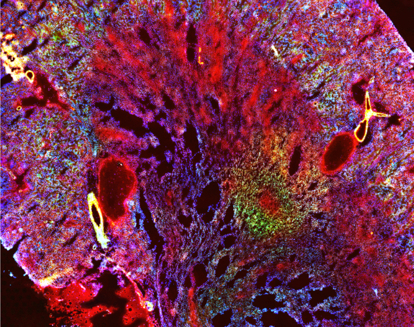
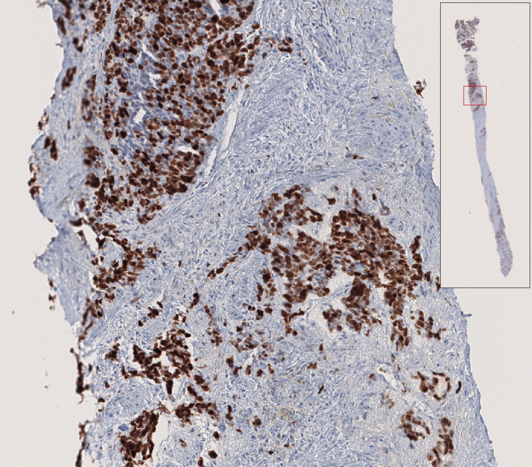
Spatial analysis of pancreatic adenocarcinoma immmunomicroenvironment
Pancreatic ductal adenocarcinoma (PDAC) has a poor prognosis, with a mortality rate exceeding 90%, making it a deadly disease. Despite advances in oncology, there has been little progress in improving the prognosis and therapy for...
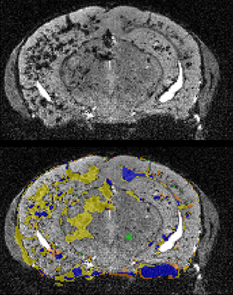
Segmentation of cavernous malformations using MRI scans of mouse brain
Cerebral Cavernous Malformations (CCM) is a vascular disease that originates in the endothelial cells lining blood vessels and which results in the formation of hallmark popcorn shaped lesions in the brain, spine, and/or...

The role of perivascular TAM phenotypes in metastatic dissemination
Accumulation of Tumor-Associated Macrophages (TAMs) is associated with enhanced tumor progression and poor survival in most solid cancers. TAMs are plastic cells exhibiting states and phenotypes spanning from anti-tumor/immunostimulating (sometimes referred to as...
Ultrastructural protein mapping through Correlation of Light and Electron Microscopy
The project focuses on accurately aligning LM and EM images to combine molecular and ultrastructural information for high-resolution analysis of endo-lysosomal alterations.
A 3D human motor neuron disease platform for high throughput drug screening
The project developed a scalable 3D screening platform for analysing human motor neuron disease (MND) models using high-content microscopy.
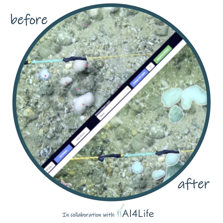
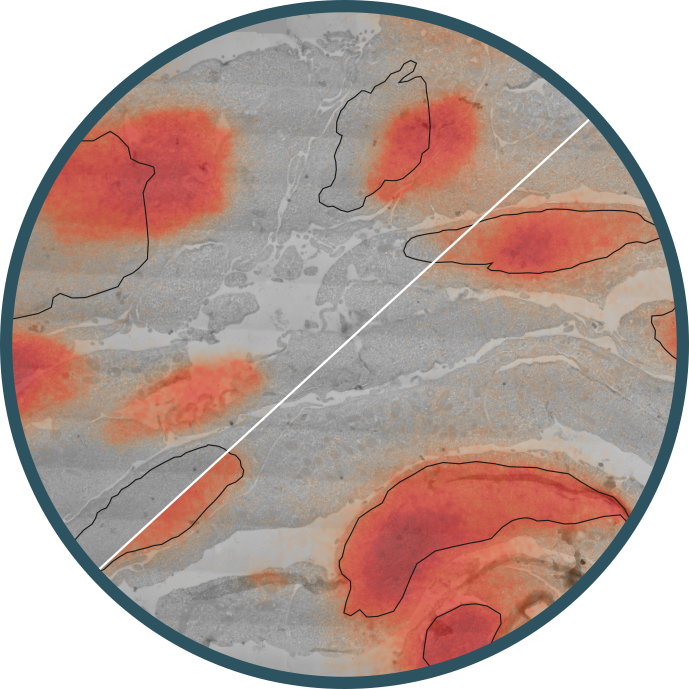
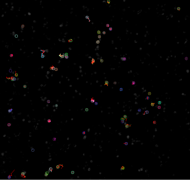
Automated species annotation in turbid waters
The project’s aim was to create an automated image-annotation pipeline to monitor the ecological status of vulnerable marine habitats according to the goals of multiple European Directives.
Particle tracking for drug diffusion and dissolution in the mucus layer
Nanoparticles used as drug carriers diffuse at different rates depending on their interactions with the surrounding medium. In the colon, this medium is colonic mucus, a complex biological barrier that strongly influences particle...
Ki-67 and PSA quantification
The aim of this project is to explore the potential of digital pathology to perform automatic scoring of the immunohistochemistry markers Ki-67 and PSA in prostate cancer biopsies.
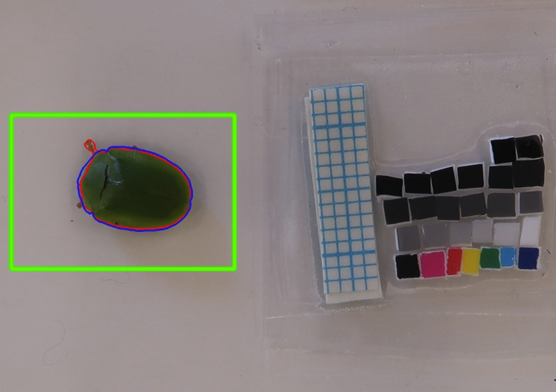
Phenotyping of tortoise beetles: Automated extraction of morphology from thousands of beetle pictures...
We want to investigate why populations differ in their ability to evolve and adapt. Our aim is to extract morphological traits, such as body size and shape, from pictures of our study species the green...
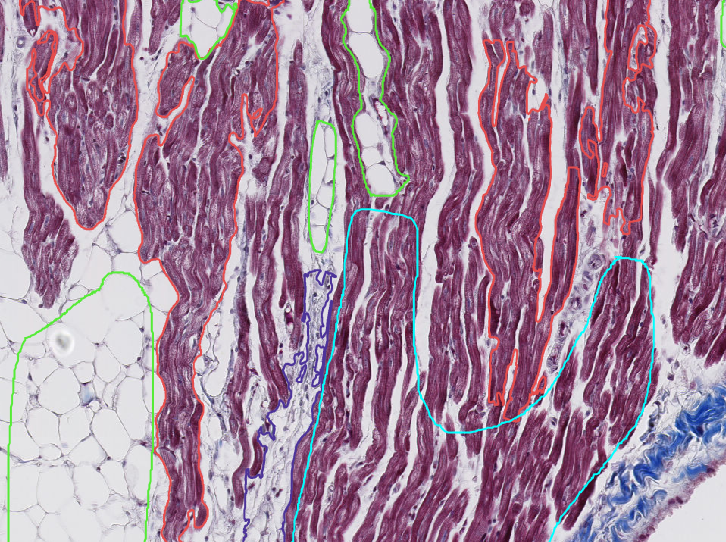
Pathological characterization of cardiac tissue samples from dogs in various stages of myxomatous...
Histopathology will be employed to characterize cardiac tissue samples collected from dogs affected by Myxomatous Mitral Valve Disease (MMVD) or Dilated Cardiomyopathy (DCM) to increase knowledge about the cardiac remodeling process in cardiac diseased dogs....
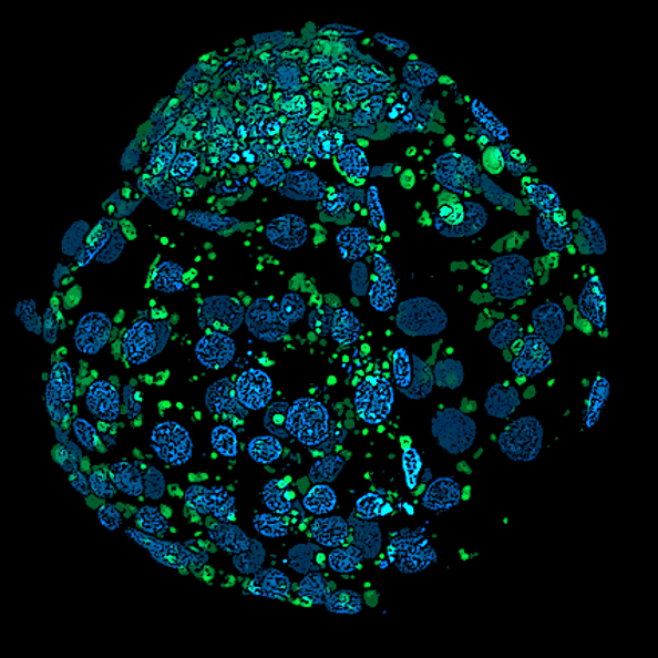
Image analysis of bovine embryos
At SLU in Uppsala, Sweden’s only in vitro embryo production laboratory for agricultural animals is located. The laboratory is well-equipped and has functional embryo production protocols for cattle and pigs. However, just like...