Illustration (click to hide):
Project Description
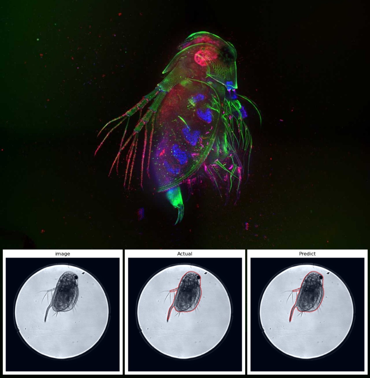
This project aims to develop an image-based analysis method for toxicological effects in Daphnia magna, an ecotoxicological standard test species, after exposure to industrial chemicals or environmental samples. Molecular staining combined with fluorescence microscopy measures adverse effects on the organism. Light intensity data is extracted from fluorescence images of daphnia exposed to a range of concentrations of a chemical/sample to quantify the effects. The main goal of this study is to generate toxicological data in a time-efficient manner. Therefore, image acquisition was done with an automated multipoint confocal high-content imaging system suitable for experiments in multi-well plates. To achieve a high-throughput method, this project also requires a time-efficient image analysis method with accurate segmentation of the daphnia based on the transmitted light image. It further requires an easy application of the segmentation mask to the corresponding fluorescence images to extract intensity data.
Project Information
-
BIIF Principal Investigators
- Christophe Avenel
External Authors
Cedric Abele, Oskar Karlsson, -
Date
2024-11-06 🠚 Current - GitHub page