Illustration (click to hide):
Project Description
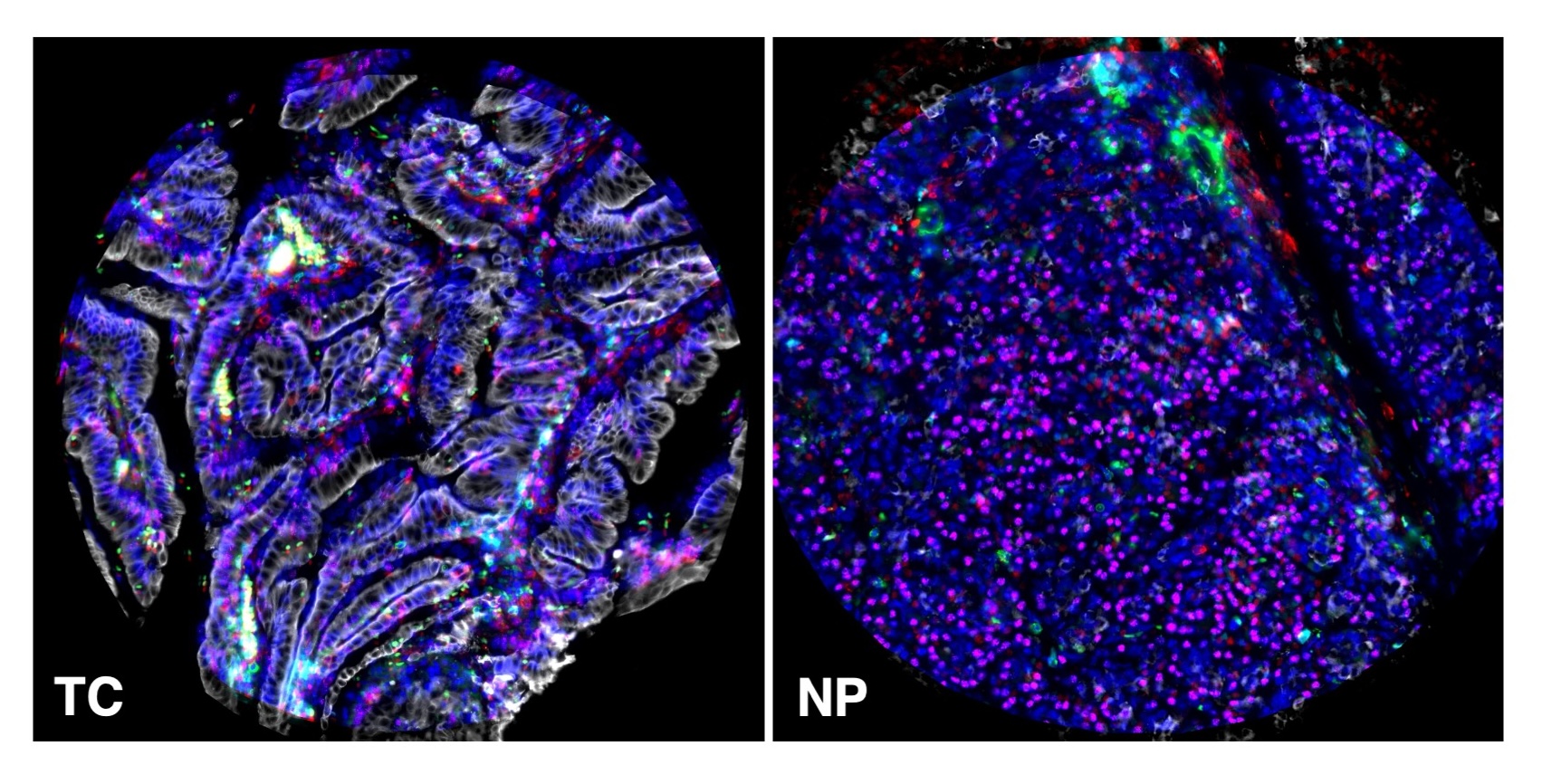
Pancreatic ductal adenocarcinoma (PDAC) has a poor prognosis, with a mortality rate exceeding 90%, making it a deadly disease. Despite advances in oncology, there has been little progress in improving the prognosis and therapy for PDAC patients. Unlike other types of cancer, where immune checkpoint inhibitors show anti-tumor efficacy, PDAC exhibits remarkable resistance. A better understanding of the PDAC immune landscape is necessary to understand the mechanisms behind this poor response to immunotherapies. Spatial transcription by 10X genomics and multiplex immunofluorescence with defined panels of immune cell subset will be used to dissect the immune microenvironments in the tumour centre, invasive front and normal parenchyma. The data collected will be evaluated in the context of the patients’ clinical response to the treatment.
Figure legend. Multiplex immunofluorescence analysis of the tumour centre (TC) and normal adjacent parenchyma (NP) of a PDAC patient. The following antibody panel was used: anti-CD8 (red), anti-CD8 (green), anti-CD79a(cyan), anti-pan cytokeratin (white), anti-TCF1 (purple).
Project Information
-
BIIF Principal Investigators
- Christophe Avenel
External Authors
Ioannis Pateras, Jean Descarpentrie, Konstantinos Ntostoglou, Teresa Frisan, -
Date
2024-09-01 🠚 Current