Illustration (click to hide):
Project Description
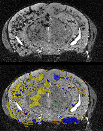
Cerebral Cavernous Malformations (CCM) is a vascular disease that originates in the endothelial cells lining blood vessels and which results in the formation of hallmark popcorn shaped lesions in the brain, spine, and/or retina. In the clinic, doctors rely on magnetic resonance imaging (MRI) of patients to view the location and functional characteristics of CCM lesions. Research groups use different mouse models in order to study CCM in the lab, and pre-clinical MRI could be an invaluable tool. This project aims to generate a user-friendly pipeline that allows for the assessment of disease burden using MRI images of mouse brain, which could also be adapted to related studies with 3D or 4D datasets in the brain.
Project Information
-
BIIF Principal Investigators
- Agustin Corbat
External Authors
Ross Smith, Peetra Magnusson, -
Date
2024-09-01 🠚 2024-12-21 - GitHub page