Illustration (click to hide):
Project Description
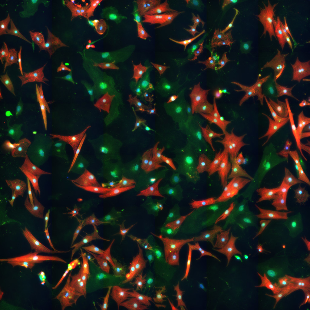
Redox homeostasis is largely dysregulated in heart failure as NADH is produced on the expenses of NADPH to support ATP production under maladaptive remodeling (Nickel, von Hardenberg et al. 2015). Moreover, remodeling in heart failure has been associated with enhanced ROS production due to activation of sympathetic nervous system and the renin–angiotensin–aldosterone system, or due to altered cytoplasmic calcium handling (Münzel, Gori et al. 2015). But despite this fact, clinical trials focusing on ameliorating oxidative stress and improving cardiac energetics have not achieved clinical success (de Zeeuw, Akizawa et al. 2013, Zhou and Tian 2018). We have noted that redox stress induces changes to cardiomyocytes geometry, where oxidative stress induces cardiomyocytes’ elongation and atrophy, while reductive stress induces cardiomyocytes hypertrophy. These alterations in the cardiomyocyte’s geometry resemble the changes observed to heart geometry during heart failure. To have quantitative measurements for these cellular alterations, we treated neonatal cardiomyocytes with a compound that induces oxidative stress (Sulforaphane) and a compound that induces reductive stress (N-Acetyl Cysteine). We aim to measure cell geometry alterations by florescence microscopy and to correlate that to molecular data obtained from the same treated cells. We anticipate applying the outcomes of this experiment in designing an in vivo study for a selective antioxidant treatment for heart failure.
Project Information
-
BIIF Principal Investigators
- Gisele Miranda
External Authors
Christer Betsholtz, Zaher Elbeck -
Date
2021-04-13 🠚 Current