Illustration (click to hide):
Project Description
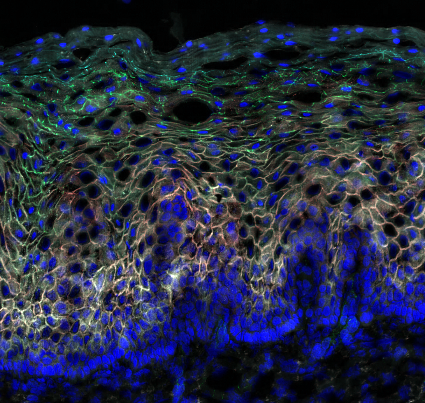
The ectocervical epithelium represents the actual first line of defense against sexual transmitted infections. To understand the quality of the protective epithelial border, we use in situ staining combined with digital image analysis to visualize and characterize the intrinsic network between, epithelial cells, immune cells and other surrounding structures the ectocervical epithelium. The ectocervical epithelium consist of a multilayered squamous epithelium where the basal membrane provides the support for the first layer of the epithelium, the basal layer. The basal layer furthermore separates the epithelium from the underlying submucosa/stroma. In our current digital image analysis pipelines we need to manually define the basal membrane (Fig 1) as a first step to define the region of interest (i.e. epithelium) within the tissue section that we want to analyze. Through collaboration with BIIF we want to see if it if is possible to design a digital analysis pipeline which allows for automatic detection of the basal membrane/layer. Such improvement would not only speed up our analysis, but it would also make sure that the separation of the epithelium form the submucosa will be done in an objective manner.
Project Information
-
BIIF Principal Investigators
- Gisele Miranda
External Authors
Annelie Tjernlund, Kristina Broliden, Gabriella Edfeldt, Mathias Boger -
Date
2019-10-25 🠚 Current