Illustration (click to hide):
Project Description
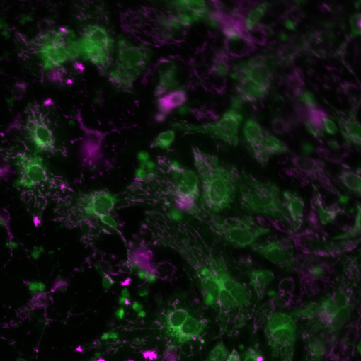
Membrane potential has two basic functions. First, it allows a cell to function as a battery, providing power to operate a variety of “molecular devices” embedded in the membrane. Second, in electrically excitable cells such as neurons and muscle cells, it is used for transmitting signals between different parts of a cell. Probes that detect mitochondrial membrane potential are positively charged, causing them to accumulate in the electronegative interior of the mitochondrion. JC-1 dye exhibits potential-dependent accumulation in mitochondria, indicated by a fluorescence emission shift from green (~529 nm) to red (~590 nm). Consequently, mitochondrial depolarization is indicated by a decrease in the red/green fluorescence intensity ratio. The potential-sensitive color shift is due to concentration-dependent formation of red fluorescent J-aggregates The aim of this project is to study if there are changes in levels of MMP or mitochondrial dysfunction in SNAT10 KO mouse cortical neurons compared to wildtype. For this we need to quantify the emission shift from green to red.
Project Information
-
BIIF Principal Investigators
- Anna Klemm
External Authors
Robert Fredriksson, Rekha Tripathi -
Date
2019-10-23 🠚 Current