Illustration (click to hide):
Project Description
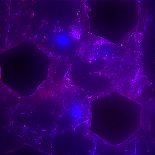
Bone defects are a common and sometimes very difficult problem in the patient population. If the body can’t heal the defect by itself, the gold standard treatment is bone grafting. This type of transplantation is the second most common worldwide, only surpassed by blood transfusion. There are several drawbacks with bone grafting: lack of availability, pain from the donor site and some immunological aspects when it comes to allografting. Biomaterials is a promising field for replacing bone and the current project is evaluating different types of 3D-printed scaffolds for bone regeneration. Bone cells (osteoblasts) derived from mouse are seeded onto these scaffolds and the cells are left to grow and produce bone matrix for four weeks. Cytoplasm and cell nucleus is then stained with Phalloidin/DAPI. Newly made bone is stained with Tetracycline that is incorporated already in the bone forming process. The image analysis will provide useful data for the quantification of cell establishment and growth.
Tags: Microscopy, model organisms and tissues
Project Information
-
BIIF Principal Investigators
- Petter Ranefall
- Carolina Wählby
External Authors
Nils Hailer, Anders Westermark, Erik Engberg, Department of Surgical Sciences, Uppsala University -
Date
2018-09-19 🠚 Current