Illustration (click to hide):
Project Description
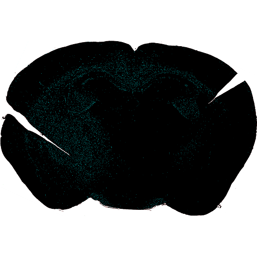
The main goal of this study was to develop a platform for quantifying the tumor-initiating capacity of a large panel of glioma-stem cell cultures (GSCs) in adult mouse brain to define the cancer stem-cell like property of the individual cultures and to integrate the result with genomic and transcriptional profiling of the GSCs. In order to achieve this, adherently grown GFP-luciferase GSC cultures were dissociated and injected stereotactically into the brain of immunodeficient mice. Tumor growth was monitored in vivo by bioluminescence imaging for up to 40 weeks and brains were collected for histopathological and immunohistochemical stainings. Automatic quantification and growth pattern analysis of tumor cells in brain sections was set up based on human cell specific staining using NuMa antibodies and a CellProfiler Analyst machine learning classifier with a manual observer correlation of 0.86. Tumors were identified in brains from mice injected with 15/29 GSC cultures, suggesting these cells as a valuable resource for future preclinical therapeutic studies targeting predicted vulnerabilities for individual glioma patients.
Tags: Microscopy, model organisms and tissues
Project Information
-
BIIF Principal Investigators
- Petter Ranefall
- Carolina Wählby
External Authors
Riasat Islam, Cecilia Krona and Sven Nelander. Dept. of Immunology, Genetics and Pathology, UU -
Date
2016-10-07 🠚 2017-03-21