Illustration (click to hide):
Project Description
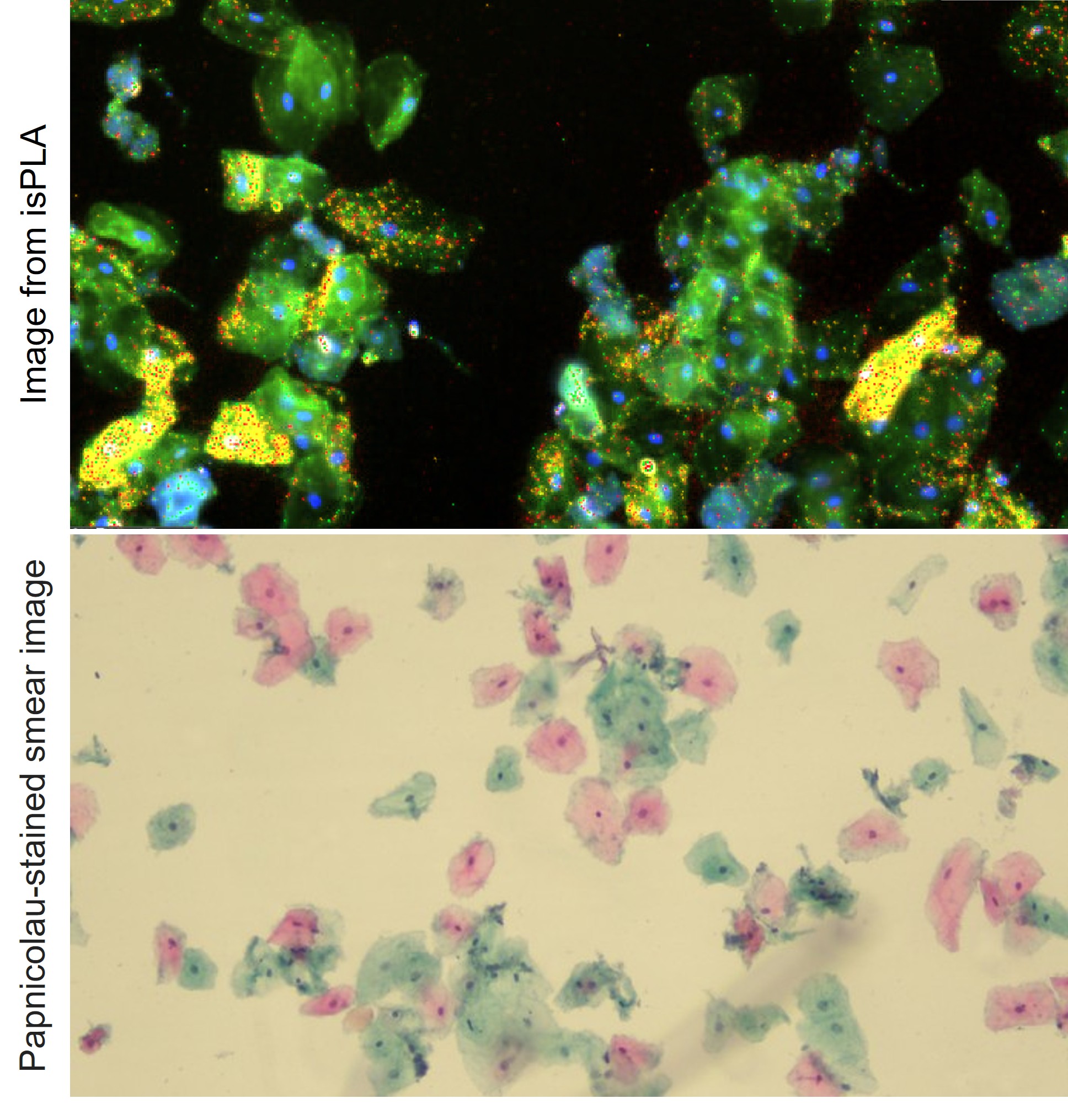
The incidence of oral cancer is increasing in the world and using a cytobrush to obtain a couple of thousand cells extracted from the abnormal area, typically located in the mucosa or gingiva, can be a cheap, non-invasive technique for evaluating cells repetitively. The cells are analyzed at both the morphological level, through visualization with standard Papanicolaou staining, and at the protein level, by staining with antibodies and applying in situ proximity ligation assay(isPLA). We aim to develop a machine learning pipeline to quantify and differentiate single cells based on the number of signals, signal intensity and provide a wider analysis of potential malignant cells.
Project Information
-
BIIF Principal Investigators
- Suganya Sivagurunathan
- Christophe Avenel
External Authors
caroline.dahlstrom@igp.uu.se, Ulf Landegren, -
Date
2025-09-18 🠚 Current - GitHub page