Illustration (click to hide):

Project Description
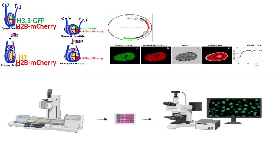
In cells of the human body, inactive heterochromatin tends to distribute in the periphery of the cell nucleus, whereas active euchromatin is mostly localized in the nuclear interior. This spatial organization essential for chromosome homeostasis and control of gene expression and is controlled by proteins in the nuclear membrane. FRIC (Fluorescent Ratiometric Imaging of Chromatin) is a quantitative image analysis method that allows monitoring of chromatin distribution in live cells. Using FRIC, it is possible to detect changes in chromatin organization resulting from silencing or overexpression of nuclear membrane proteins or treatment with epigenetic drugs. In order to perform systematic screening siRNA libraries targeting all annotated nuclear membrane proteins in a cell type, FRIC must be implemented for high content screening (HCS) conditions. This requires further development of the FRIC method to enable automated image sampling and image segmentation. Using HCS-FRIC, we expect to identify novel nuclear membrane proteins involved in chromatin organization and small organic substances able to correct disorganized chromatin in various diseases as epigenetic drug candidates.
Project Information
-
BIIF Principal Investigators
- Jonas Windhager
External Authors
Einar Hallberg, Urška Kašnik, Saira Aftab -
Date
2023-08-09 🠚 Current