Illustration (click to hide):
Project Description
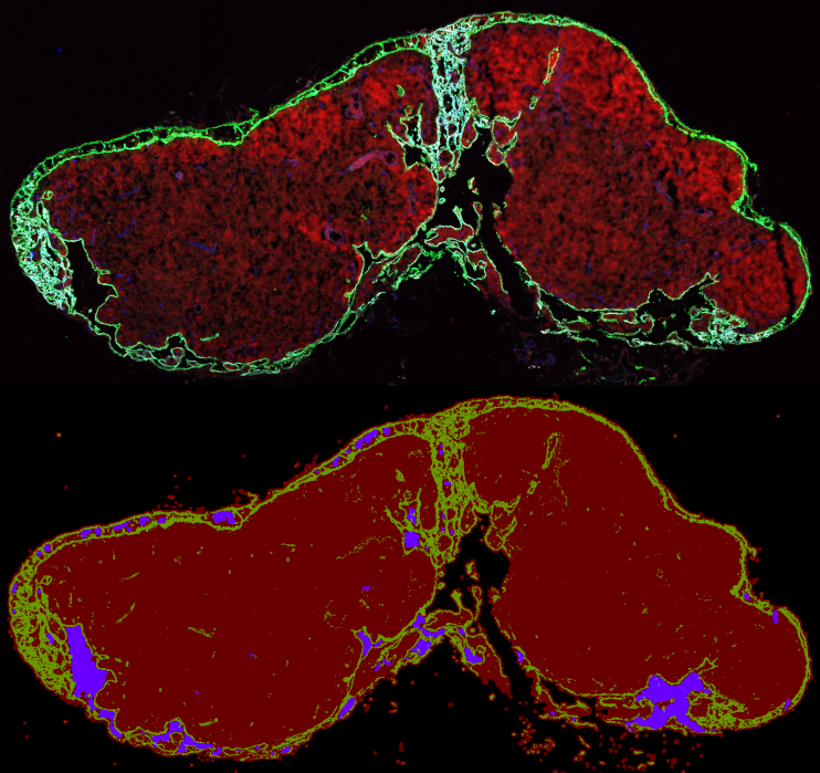
The aim of the project is to create an ImageJ macro that can be used to measure three different areas in confocal images of tissue sections; (1) the whole section area, (2) the vessel area and (3) the lumen area within the vessels. The researcher provides a .lif file with merged tilescan images in four channels: Channel 1: DAPI stain; Channel 2: GFP reporter; Channel 3: LYVE1 stain and Channel 4: VEGFR2 stain. An overlay of all four channels is used to measure the total area. From the overlay of all four channels, all the lumens (=areas with no staining) that are located within the vessels (Channels 3-4) is measured. An overlay of channels 3-4 are used to measure the vessel area. The macro creates a result folder containing a .csv file with the measurements, a TIF file with the maximum projection image and a TIF file with the masked measurement areas in RGB (see Figure above).
Project Information
-
BIIF Principal Investigators
- Christophe Avenel
- Petter Ranefall
External Authors
Bojana Jakic, Taija Mäkinen -
Date
2020-09-07 🠚 2020-09-18