Illustration (click to hide):
Project Description
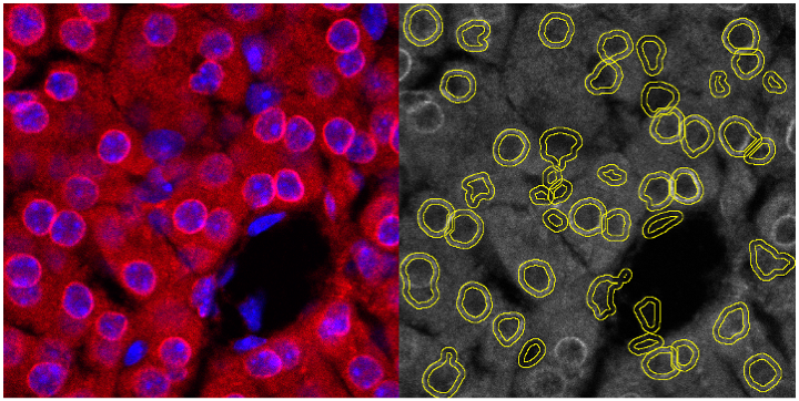
The Hutchinson-Gilford progeria syndrome is a very rare segmental progeroid syndrome, affecting children at an early age. It is characterized by clinical features resembling that of marked premature aging, including alopecia, scleroderma-like skin changes, loss of subcutaneous adipose tissue, skeletal dysplasia and cardiovascular disorders, the latter leading to premature death at an average age of 14.6 years. This disease is caused by a de novo point mutation in the LMNA gene (c.1824 C>T), which leads to the activation of a cryptic splice site and alters the normal processing of the lamin A protein, a nuclear lamina protein playing multiple important roles at the cellular level. Lamin A is first synthesized as a precursor protein termed prelamin A, which undergoes several post-translational modifications before reaching its mature form. In the case of HGPS, prelamin A appears as truncated and cannot be normally processed, which results in the production of a toxic protein called progerin, which accumulates at the nuclear rim and disrupts the nuclear shape and functions. Several animal models have been developed to mimic HGPS common features, among which the Zmpste24-/- (which accumulates prelamin A) and LmnaG609G (which accumulates the murine form of progerin) mice are the most predominantly used models. Various treatment strategies have emerged since the discovery of the mutation leading to HGPS, of which many of them are assessed in in vivo models of HGPS. To understand how treatments may improve the health- and lifespan of progeroid mice, we assessed the nuclear distribution of various nuclear proteins that play a critical role in the pathophysiology of HGPS, and in particular the distribution of both prelamin A and progerin in Zmpste24-/- and LmnaG609G mice cells. We performed immunofluorescent stainings against those proteins and assessed their relocalization at the nucleoplasm compared to the nuclear rim by image analysis and assessment of the fluorescence intensity. Relocalization of those proteins may improve the nuclear functions, in turn contributing to an improvement of the HGPS phenotypes commonly observed in those mice.
Project Information
-
BIIF Principal Investigators
- Gisele Miranda
External Authors
Maria Eriksson, Gwladys Revechon -
Date
2019-07-19 🠚 2019-09-05