Illustration (click to hide):
Project Description
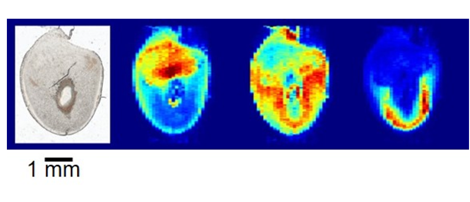
Mass spectrometry imaging enables visualization of the chemical microenvironment in regional features within thin tissue sections. In this project, thin tissue sections of mouse embryo implantation sites on day 8 of pregnancy were imaged with nanospray desorption electrospray ionization (nano-DESI) mass spectrometry to reveal molecular signatures during embryonic development. Nano-DESI is a recently established non-commerially availabe technique for mass spectrometry imaging that yields chemical information from localizations on the tissue without the need for sample preparation. The acquired data contains a full mass spectrum, with thousands of peaks, in each pixel of the image in a format that is currently not readable by commerial softwares. The aim of the project is to streamline data processing and analysis to find co-localizing and anti-localizing molecules to further our understanding of the importance of molecular localization in successful pregnancy. In particular, tissue sections from wild type and knock out mice, that deliver pups prematurely, will be evaluated. A successful project is anticipated to provide novel image analysis tools for data collected by nano-DESI mass spectrometry imaging of biological systems.
Tags: Microscopy, model organisms and tissues
Project Information
-
BIIF Principal Investigators
- Anna Klemm
- Carolina Wählby
External Authors
Ingela Lanekoff, Uppsala Univeristy -
Date
2018-06-28 🠚 2018-08-31