Illustration (click to hide):
Project Description
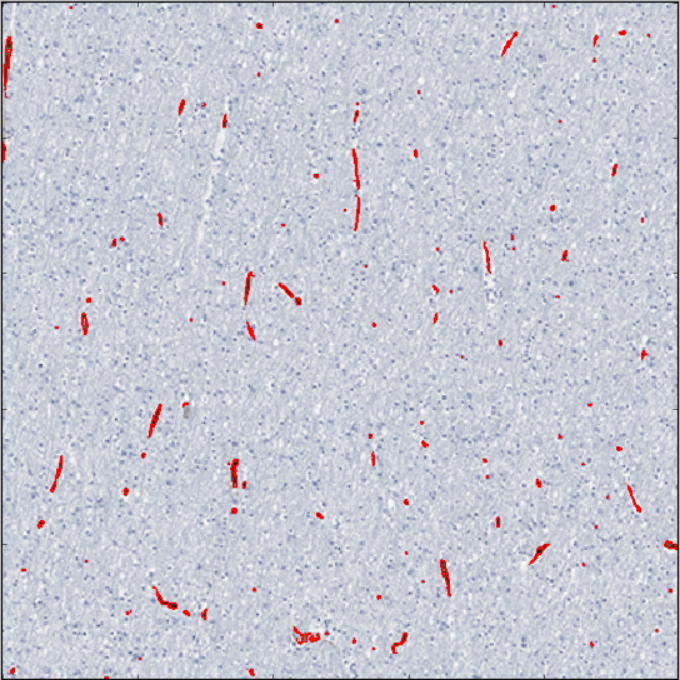
Gliomas are heterogeneous tumors in terms of imaging appearances, and a deeper understanding of the histopathological tumor characteristics that underlie the signal abnormalities on PET and MRI is needed. Here we used histology-to-radiology co-registration of gliomas with the aim to correlate local changes in tumor perfusion and 11C-methionine uptake with cell density, vascularity and proliferation in these areas.
Tags: Microscopy, cell biology
Project Information
-
BIIF Principal Investigators
- Petter Ranefall
External Authors
Kenney Roodakker, Anja Smits, Department of Neurology, Uppsala University -
Date
2017-11-30 🠚 2018-12-31